
Graphene Microtransistors for the mapping of Brain Activity: An Article Review
Recently, fluctuations of brain activity occurring at less than 0.1 Hz has been a center of interest. These fluctuations less than 0.1 Hz are commonly referred as very slow, ultraslow and infraslow activity (ISA). ISA is an indicative of brain states such as sleep, coma, anaesthesia, wakefulness, etc. Cortical spreading depression (CSD), which is a slowly propagating wave of near-complete depolarization of neurons, occurs at infralow frequencies. CSD generally arises from stroke or brain injury as well as migraines. Therefore, monitoring these signals can be vital for clinical diagnosis and therapies. To record these signals, microelectrodes are used, but current microelectrode materials limit this recording process because of voltage drift and high electrode impedance. To prevent these poor conditions, high-pass filters are used, but it also has some disadvantages which is loss of physiological and pathological information.
Disclaimer: The content of this post or any other linked material is intended for informational purposes only and should not be taken as medical or technical advice.
A group of researchers from Spain have found a solution for these problems explained in detail in their article “High-resolution mapping of infraslow cortical brain activity enabled by graphene microtransistors” published in Nature Materials. They used flexible epicortical and intracortical arrays of graphene solution-gated field-effect transistors (gSGFETs) in rats. Results were satisfying, gSGFETs were able to record infraslow signals together with other typical signals with high accuracy as can be understood from the Figure 1. The reason why gSGFETs were successful is that graphene provides a great flexibility which helps gSGFETs to be work with ultra-soft and flexible substrate without reduction in performance. Moreover, graphene does not present a threat in clinic applications due to its high biocompatibility and excellent electrochemical stability.
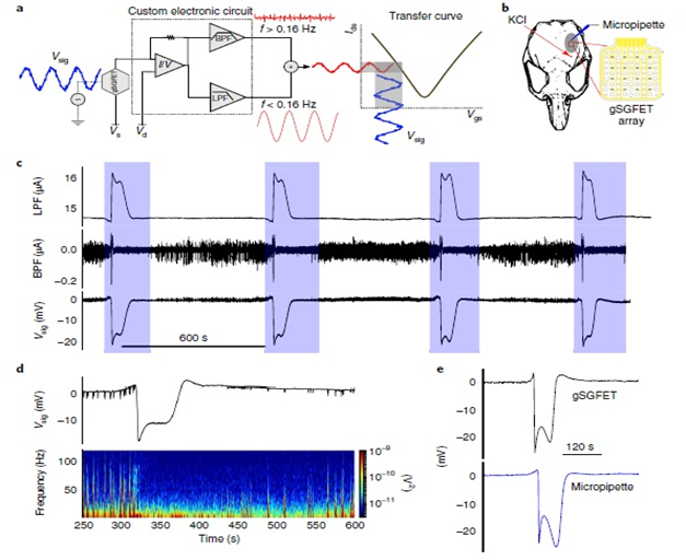
Figure 1: a) Schematic of the gSGFET recording setup. b) Schematic of a rat skull depicting the craniotomy (grey area). c) Electrophysiological recordings obtained with a gSGFET epicortical array during the induction of four CSD events (blue shade). d) Voltage-converted wide-band signal of a CSD event recorded by a gSGFET and spectrogram showing the characteristic silencing of activity. e) Comparison of a CSD signal recorded by a graphene transistor and solution-filled glass micropipette with a Ag/AgCl wire demonstrating the excellent similarity in shape, magnitude and time span.
In conclusion, it has been shown that gSGFETs can record signals from infralow (< 0.1 Hz) to the typical local field potential bandwidth. They outperform solution-filled glass micropipettes by overcoming their spatial sampling limitations. Also, gSGFETs provide convenience to their application by enabling design of wide range of device sizes and they give high accuracy and spatial resolution in mapping of infralow oscillations. All these properties make the understanding brain regions where ISA is initiated. Therefore, it is seen that this technology will lead to a better understanding in clinic and laboratory research.
Read: Ultimate Guide to Graphene: Everything You Need to Know About Graphene

Recent Posts
-
 in Food Industry")
Cellulose Nanocrystals (CNC) in Food Industry
Cellulose nanocrystals (CNC) are emerging as a pivotal material in the realm of food technology, her …3rd May 2024 -

Reducing the Carbon Footprint of Nanomaterials
The production of nanomaterials is vital for numerous advanced applications, from healthcare to elec …26th Apr 2024 -

Nanocomposites in Food Packaging
The utilization of nanocomposites in food packaging represents a significant advancement in the fiel …19th Apr 2024