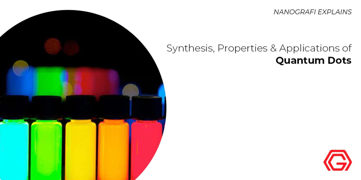
Synthesis, Properties and Applications of Quantum Dots
Quantum dots (QDs) are generally fluorescent semiconductor nanoparticles sized less than 20 nm with each particle containing some 200 to 1000 atoms.
Depending on a phenomenon called “quantum confinement” and their surface to volume ratio, quantum dots adopt particular electronic and optical properties.
Vahid Javan Kouzegaran
Analytical Chemist (Ph.D.) / Nanografi Nano Technology
Introduction
Quantum dots structure is made of two main parts as a core which is protected by a shell. The shell is made of ZnO, ZnSe, PbS, ZnTe, CdSe, CdS and compounds that can protect the core from any degrading reactions and more importantly maintain fluorescence properties. The core in quantum dots are usually made of semiconducting phosphides (InP and GaP) chalcogenides of lead, cadmium, zinc (CdSe, CdTe and DdS), nitrides (GaN) arsenides (InAs and GaAs) and Copper salts 1.
Synthesis of Quantum Dots
Similar to two common methods used to synthesize nanoparticles, quantum dots are prepared through bottom-up and top-down synthetic techniques. Based the top-down method which is originally physical, a bulk semiconductor material is thinned using ion implantation, e-beam lithography, X-ray lithography, molecular beam epitaxy (MBE). The bottom-up approach, however, is through a chemical reaction in aqueous environment which itself involves two main methods known as vapor-phase and wet-chemical methods. Wet-chemical methods include hot-solution decomposition, microemulsion, sol-gel, electrochemistry, microwaves and sonic waves. Accordingly, the wet-chemical method is following the known precipitation reactions through which the significant factors as a combination of solutions are controlled to carefully control the particle size. Additionally, the method includes limited growth of particles and nucleation. Through nucleation, particles of uniform sizes (homogeneity) or different sizes (heterogeneity) are obtained 2.
Properties of Quantum Dots
Quantum dots are regarded as nanoscale colloidal single crystal semiconductors whose size and morphology can accurately be controlled during the synthesis process through optimizing chemical factors like pH, temperature and reagents concentration. The fluorescence quality of quantum dots starts with formation of an electron-hole pair known as exciton induced by the absorption of a photon with the energy above the bang gap of the semiconductor. Based on this, quantum dots are employed as highly efficient probes for two-photon confocal microscopy. These fluorescent nanoparticles can be used simultaneously with dyes for potential immunofluorescence purposes generating higher fluorescent quantum yield and intensity as well as low toxicity 3.
Since quantum dots are synthesized in nonpolar organic solvents in most cases and their applications in polar solutions are also required, some strategies have been developed in order to increase their solubility. Replacing ligands with simple thiolated molecules, peptides, dendrons and oligomeric phosphines is among the most common functionalization techniques towards a desired solubility. Other functionalization methods are embedding them by triblock copolymers and polymer shells, silica shells and amphiphilic polysaccharides as well as combining QDs in layers of molecules 4.
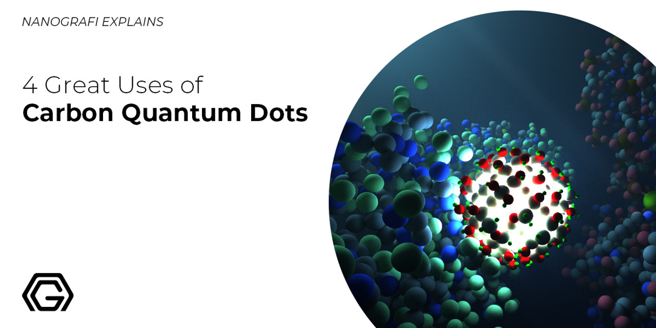
To learn more about great uses of Carbon Quantum Dots,
you can read our blog post here.
Applications of Quantum Dots
Due to the unique nanoscale along with luminescence properties, quantum dots have found a lot of applications in biosensing, gene and drug delivery, therapy and bioimaging. Below, brief descriptions will be presented concerning the applications mentioned above.
Quantum Dots as Biosensors
There has been intensive development of Forster resonance energy transfer (FRET) and fluorescence-based biosensors using quantum dots as ultrasensitive detection probes of biomolecules such as sugars, antibodies, antigens, acids and enzymes. These detections take QDs narrow emission band advantage making the detection of G-quadruplex DNAs antibodies, proteins and toxins possible. QDs can be assembled with organic dyes such as fluorescein to be used as pH or FRET sensors. They can be coupled with a variety of anticancer antibodies to detect cancer biomarkers in immunochromatographs or immune-microchannels. Quantum dots conjugation with nucleic acids and serve as efficient FRET platforms to detect dye-labeled complementary strands. This is in part similar to using gold nanoparticles which is based on surface plasmon to detect oligonucleotides. The distance between the quantum dots and dyes is the significant point in achieving a high sensitivity in these detections. In this order, preparing biosensors made of quantum dots with decent hydrodynamic diameter requires a carful setting of the distance between donor and acceptor agents 5.
Gene and Drug Delivery
In gene and drug delivery, quantum dots are functionalized to be employed as platforms to carry genes and drugs. In order to transport siRNA as remarkable transportation application, siRNA is adsorbed on the functionalized quantum dots through incubation and based on the proton sponge effect 5. In a study, fluorescence property of quantum dots has been used to facilitate intercellular delivery and transport genes and drugs through incorporating with liposomes hydrophilic cavity and a series of fragments like folic acid, membrane antigen and aptamers. In another research, imine conjugated quantum dots have been used for intracellular delivery of genes and drugs facilitating cargo release.
Therapy
In photodynamic treatment, quantum dots are studied solely or with photosensitizers including inorganic complexes, porphyries and organic dyes for the purpose of cancer imaging and delivering therapeutics to increase cancer therapy quality. In so doing, singlet oxygen and other oxygen compounds are generated in a photosynthetic process. Later, quantum dots as extremely photostable agents activate singlet oxygens in the therapy. Peptides, amines or carboxylic acids engage in conjugating sensitizing singlet oxygen quantum dots. In this way, antibodies can locate quantum dots in cancer cell or tumors 6.
Bioimaging
Actually, quantum dots are mainly used for the purpose of cell imaging. The application of quantum dots in this context rely on properties of molecules immobilized on the surface of quantum dots. Quantum dots have the possibility to be encapsulated within small molecules or be conjugated to proteins, polymers, lysozyme carbohydrates cationic lipids and moreover, they can attach to cell membrane nonspecifically. Conjugation of quantum dots with peptides is a technique for nucleus and mitochondria intracellular imaging and labeling. Using liposomes and surfactants, the transportation efficiency in quantum dots cytosol increases. In contrast to the nonspecific extracellular and intracellular labeling employed to detect and image cells, specific cell biomarker are applied for detecting and imaging cancer cells and genes and for drug delivery as well. In conjugation with antibodies (anti-EGFR & anti-HER2), transferrin and growth hormones (EGF and VEGF), quantum dots are used as cancer cell imaging and detecting 7.
In addition to the interesting properties of nanoparticles, quantum dots are among the very rare nanoscale particles which are originally fluorescent without being functionalized. This quality is added to its semiconducting property making it a potential biosensing and bioimaging platform for medical purposes.
To discover the latest news from nanotechnology, you can visit Blografi.
References
1. Pantapasis, K., Anton, G. & Bontas, D. Chapter 2 - Bioengineered nanomaterials for chemotherapy. Nanostructures for Cancer Therapy (Elsevier Inc., 2017). doi:10.1016/B978-0-323-46144-3/00002-7
2. Kesharwani, P. et al. PAMAM dendrimers as promising nanocarriers for RNAi therapeutics. Mater. Today 18, 565–572 (2015).
3. Yang, H. Targeted nanosystems: Advances in targeted dendrimers for cancer therapy. Nanomedicine Nanotechnology, Biol. Med. 12, 309–316 (2016).
4. Kesharwani, P., Gajbhiye, V. & Jain, N. K. A review of nanocarriers for the delivery of small interfering RNA. Biomaterials 33, 7138–7150 (2012).
5. Biju, V. Chemical modifications and bioconjugate reactions of nanomaterials for sensing, imaging, drug delivery and therapy. Chem. Soc. Rev. 43, 744–764 (2014).
6. Park, B. K. et al. New Generation of Multifunctional Nanoparticles for Cancer Imaging and Therapy. 1553–1566 (2009). doi:10.1002/adfm.200801655
7. Frigerio, C. et al. Analytica Chimica Acta Application of quantum dots as analytical tools in automated chemical analysis : A review. Anal. Chim. Acta 735, 9–22 (2012).
Recent Posts
-
Reducing the Carbon Footprint of Nanomaterials
The production of nanomaterials is vital for numerous advanced applications, from healthcare to elec …26th Apr 2024 -
Nanocomposites in Food Packaging
The utilization of nanocomposites in food packaging represents a significant advancement in the fiel …19th Apr 2024 -
What is the Difference Between 7075 and 6061 Aluminum Alloy?
When comparing 7075 aluminum alloy to 6061 aluminum alloy, it's essential to understand their disti …5th Apr 2024


