Gold Nanostars - Nanoparticles - Nanografi Blog
Gold Nanostars are anisotropic gold nanoparticles. Their structure is composed of a core and several sharp branches. The unique properties of gold nanostars stem from these branches and their interaction with the core. The most attractive features of gold nanostars are the LSPR, SERS activity, and catalytic activity. Gold nanostars are used in catalysis, nanosensing assays, thermal therapy, and drug delivery applications.
Click Image to Reach Our Other Articles About Health and Delivery Systems
The improvements in nanotechnology have been a great success in different areas of science. As the scientists succeeded to obtain nano-sized structures and discovered the powerful properties of these structures, they have thrived to come up with improved versions in the nanosized realm. Much like many other areas of science, nanotechnology takes its inspiration from nature. The hierarchical structure of marine starfishes and sea urchins has been the foundation of new nanosized materials. These new structures have multiple rays of rams extending through a core body which increases the surface to volume ratio, active surfaces, and adsorptive surfaces. One of the important members of these novel structures is star-shaped nanoparticles known as nanostars. Most of the researches on nanostars mainly focuses on gold nanostars even though some examples of palladium, platinum, silver, and rhodium are also present in the literature (1).
Gold nanostars are a member of anisotropic gold nanoparticle category. They attract particular interest due to their shape modifications, such as the number of branches, aspect ratio, and length of branches (2).
Fabrication of Gold Nanostars
The synthesis of gold nanostars is mostly based on the chemical reduction of gold salts. There are two different approaches to gold nanostar fabrication. The first approach is known as the seeded-growth method. In the seeded-growth technique preformed seeds act as nucleation points and the additional materials deposit on these seeds. This method usually leads to monodisperse particles with different sizes. This method involves the reduction of chloroauric acid (HAuCl4) with ascorbic acid at room temperature, on pre-synthesized gold nanocrystal seeds and in the presence of a cationic surfactant. These surfactants are also known as capping agents and believed to trigger the anisotropic growth process. However, the use of these agents requires post-treatments for separation from the gold nanostar structure. The nature of seeds and reducing agents are the important parameters for the morphology of resulting gold nanostars. Frequently used capping agents are; typically cetyl-trimethylammonium bromide, CTAB), cetyl-trimethylammonium chloride (CTAC), N, N-dimethylformamide (DMF) and hydroxylamine sulfate (4).
Click Image to Check Another Article About Gold Nanostars
On the other hand, in one pot methods in situ formation of seeds are observed. Then the subsequent growth of branches takes place. This method usually involves the use of reducing agents and surfactants under room temperature and verity of reactant concentrations and reaction times. For this method, frequently used surfactants are CTAB, SDS, and PVP while reducing agents are ascorbic acid, hydrazine, and DMF. However, through the one-pot method, it is also possible to prepare gold nanostars without the use of capping agents. The absence of capping agents reduces the complexity of post-separation steps. Even though this method is highly sensitive to synthetic changes resulting in a difficult control of sizes and morphologies, their use has widely spread due to their highversatility (1).
Properties of Gold Nanostars
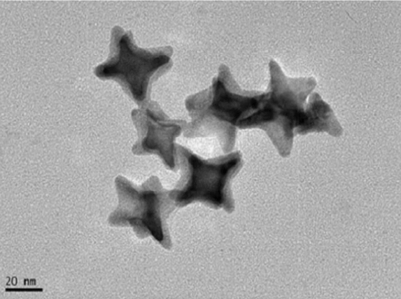
The morphology of gold nanostars ranges from a simple three-branched structure to multiple sharp branches. As the nanostar size increases, the core size, the number of branches, and the branch aspect ratio also increase. With the increasing size, the stars become more inhomogenous in shape while the overall size remains homogenous. The proximity, size, and the number of branches affect the properties of gold nanostars. The main properties that have attracted considerable attention to gold nanostars are their optical properties such as localized surface plasmon resonances (LSPR) and surface-enhanced Raman scattering (SERS), as well as catalytic properties. The intrinsic optical properties of gold nanostars result from the hybridization of plasmons focalized at the core and the tips of the nanoparticles. The core of gold nanostars acts as an antenna producing electromagnetic field enhancements of the tip plasmons. The Plasmon frequency and intensity of gold nanostars depend on the arrangement and the number of spikes. The morphology of spikes is controlled through different fabrication conditions. The unique nature of the gold nanostars displays strongly localized surface plasmon resonances (LSPRs) which are related to collective oscillations of conduction electrons produced during interaction of the metal with visible or near-IR light. Relatively lower LSPRs, around 600nm, represent the core of gold nanostars while higher LSPR values are related to the core-tip interactions. By controlling the aspect ratio in the synthetic conditions, the LSPR absorption maximum of the most abundant asymmetric GNS can be positioned in the 750-1150 NIR range (3). The LSPR features of gold nanostars can even be tailored after synthesis by introducing a small amount of reducing agent to its environment. The morphology of the gold nanostar starts to change upon this introduction and allows the fine-tuning of LSPR.
Another important optical property of gold nanostars is SERS activity. The Raman enhancement is the combined effect of the local increase in electric field when the molecule is illuminated by light at its resonant wavelengthand the increase of the strength of the Raman dipole-emission transition at a longer wavelength. The electromagnetic enhancement due to LSPRs is a major contributor to the phenomena of SERS. Gold nanostars show significantly high SERS activity compared to a sphere dimer which is considered to be the paradigm SERS substrate. The enhanced electric field is strongly observed at the tips of the gold nanostars which facilitate the amplification of Raman signal. However, it is also possible to observe these enhanced electric fields between aggregated nanostars. The large Raman signal amplification allows the observation of SERS from molecules adsorbed ontogold nanostars (4).
Traditional sphere-like gold nanoparticles have been used as a valuable catalytic material for various different reactions. As of recently, anisotropic gold nanoparticles such as gold nanostars have also attracted attention for catalytic applications. The increased surface area of gold nanostars due to their several sharp tips promotes their catalytic activity (3). Furthermore, the tunable morphology of gold nanostars is expected to promote selectivity. Due to their interesting optical properties, gold nanostars also show promising photocatalytic properties.
Click Image to Check Our Advanced Materials for COVID-19
Application Areas of Gold Nanostars
Due to the above mentioned properties, gold nanostars are used in different application areas such as heterogeneous catalysis, and biomedical applications such as; nanosensing assays, thermal treatments, clinical chemistry, and delivery systems.
Nanosensing Assays
The strong LSPR properties and the accompanying SERS signals are the basis of gold nanostar applications in highly sensitive assays. In particular, SERS provides a promising method for the detection of various biomarkers (DNA, RNA, protein, etc.) due to its high sensitivity, specificity, and capability for multiple analyte detection. In nanosensing assays, gold nanostars are usually used as the SERS substrate andfacilitate the chemical sensing of analytes. The analytes can be physically or chemically absorbed on to the nanostars’ surface for detection. Absorbed analytes induce reproducible SERS enhancement factors which are a great basis for sensing applications. The SERS nanosensors can be used in various applications including pH sensing, protein detection, and gene diagnostics (3).
Thermal Therapy
Thermal therapy is a type of medical treatment in which body tissue is exposed to high temperatures (up to 45–47 °C). It is used for cancer treatment by damaging or killing the cancer cells. The spherical gold nanoparticles are not efficient for this application since they absorb visible light and the penetration of visible or UV light in tissues is limited. The optimal wavelength for best tissue penetration is ~800 nm (NIR). The tunable LSPR of gold nanostars in the NIR region makes them great candidates for thermal therapy applications. Furthermore, the use of gold nanostars reduces the damaging of non-cancerous cells.
Thermal therapy is also very effective in antimicrobial treatment. For example, the adjusted LSPR of gold nanostars in the 700–1100 nm range is found to be effective on Staphylococcus aureus biofilms (6).
Delivery Systems
The traditional medical treatment applications are in need of improvement. Scientists have been developing targeted drug delivery systems for much more efficient therapeutic applications. Due to their tunable characteristics such as size, stability, and biocompatibility; gold nanoparticles are considered as good candidates for targeted drug delivery. Gold nanostars are one of the promising gold nanoparticles because of their increased surface area, tunable size, and surface morphology. Gold nanostars are functionalized with drug molecules for enhanced therapeutic applications. The drug release from the functionalized gold nanostars is obtained by the desorption of drug molecules through ligand-exchange processes. Gold nanostars can be used to deliver drugs directly into the nucleus of a cancer cell (5).
Heterogeneous Catalysis
As it was mentioned before, the catalytic properties of gold are further enhanced in the form of gold nanostars. Gold nanostars can be used for the reduction of aromatic nitro compounds, namely 4-nitrophenol (4-NP), 4-nitroaniline (4-NA), and 4-nitrothiophenol (4-NTP). The increased catalytic activity of gold nanostars can be linked to the corners, stepped surfaces, and high electron density at their tips (2). The optical properties of gold nanostars make them great candidates for photocatalytic applications since they can efficiently harvest light and enhance photochemical transformations. Another attractive feature of gold nanostars is their tunable surface plasmon resonance band. In addition to photocatalysis, gold nanostars are also used in the electrocatalysis for methanol oxidation and oxygen reduction from the electroreduction process of H2O2 (1).
Gold nanostars are anisotropic gold nanoparticles composed of a core and several branches. Localized surface plasmon resonances (LSPR), surface-enhanced Raman scattering (SERS), and catalytic nature of gold nanostars are the most important properties for their practical applications. The catalytic properties of gold nanostars are utilized in organic reactions while the optical properties are mainly utilized in biomedical applications such as nanosensing assays, thermal therapy, and targeted drug delivery applications. Still, gold nanostars are at their infancy and have a lot more to offer to science.
References
1.Guerrero-Martínez, A., Barbosa, S., Pastoriza-Santos, I., & Liz-Marzán, L. M. (2011). Nanostars shine bright for you: colloidal synthesis, properties and applications of branched metallic nanoparticles. Current Opinion in Colloid & Interface Science, 16(2), 118-127.
2.Priecel, P., Salami, H. A., Padilla, R. H., Zhong, Z., & Lopez-Sanchez, J. A. (2016). Anisotropic gold nanoparticles: Preparation and applications in catalysis. Chinese Journal of Catalysis, 37(10), 1619-1650.
3.Khoury, C. G., & Vo-Dinh, T. (2008). Gold nanostars for surface-enhanced Raman scattering: synthesis, characterization and optimization. The Journal of Physical Chemistry C, 112(48), 18849-18859.
4.Chirico, G., Borzenkov, M., & Pallavicini, P. (2015). Gold nanostars: synthesis, properties and biomedical application. Berlin, Germany: Springer.
5.Dam, D. H. M., Lee, J. H., Sisco, P. N., Co, D. T., Zhang, M., Wasielewski, M. R., & Odom, T. W. (2012). Direct observation of nanoparticle–cancer cell nucleus interactions. ACS nano, 6(4), 3318-3326.
6.Pallavicini, P., Dona, A., Taglietti, A., Minzioni, P., Patrini, M., Dacarro, G., ... & Scarabelli, L. (2014). Self-assembled monolayers of gold nanostars: a convenient tool for near-IR photothermal biofilm eradication. Chemical Communications, 50(16), 1969-1971.
Recent Posts
-
What is the Difference Between 7075 and 6061 Aluminum Alloy?
When comparing 7075 aluminum alloy to 6061 aluminum alloy, it's essential to understand their disti …5th Apr 2024 -
Iron-Air Batteries: The Ultimate Guide
Iron-air batteries represent a significant breakthrough in energy storage technology, offering a sus …29th Mar 2024 -
Discovering the Power of 2D Materials
In material science, the discovery of two-dimensional (2D) materials represents a transformative de …22nd Mar 2024

